pDsRed2-Nuc 载体
| 质粒类型: | 荧光蛋白报告载体 |
|---|---|
| 启动子: | CMV |
| 克隆方法: | 多克隆位点,限制性内切酶 |
| 载体抗性: | Kanamycin (卡那霉素) |
| 筛选标记: | Neomycin(新霉素) |
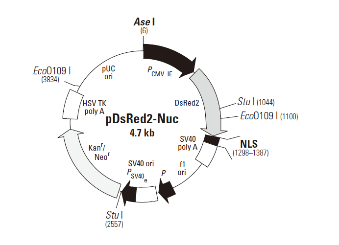
pDsRed2-Nuc is a mammalian expression vector that encodes Discosoma sp. red fluorescent protein (DsRed2; 1, 2) fused with three copies of the nuclear localization signal (NLS) of the simian virus 40 large T-antigen (3, 4). The NLS sequences are fused to the 3'-end of DsRed2—a human codon-optimized DsRed variant engineered for faster maturation and lower non-specific aggregation (1, 5). The reiteration of the NLS sequence significantly increases the efficiency with which the DsRed2 fusion translocates into the nucleus of mammalian cells (6).
To drive expression of the DsRed2 fusion, this vector contains the immediate early promoter of cytomegalovirus (PCMV IE). SV40 polyadenylation signals downstream of the DsRed2 gene direct proper processing of the 3'-end of the DsRed2-NLS mRNA transcript. To further increase the translational efficiency of the DsRed2 fusion, upstream sequences have been converted to a Kozak consensus translation initiation site (7). The vector also contains an SV40 origin for replication in any mammalian cell line that expresses the SV40 T-antigen, a pUC origin of replication for propagation in E. coli, and an f1 origin for single-stranded DNA production. A neomycin resistance cassette—consisting of the SV40 early promoter (PSV40e), the neomycin/kanamycin resistance gene of Tn5 (Neor/Kanr), and polyadenylation signals from the herpes simplex virus thymidine kinase (HSV TK poly A) gene—allows stably transfected eukaryotic cells to be selected using G418 (8). A bacterial promoter (P) upstream of this cassette drives expression of the gene encoding kanamycin resistance in E. coli.
载体应用
pDsRed2-Nuc is designed for fluorescent labeling of the nucleus in living cells. The vector can be introduced into mammalian cells using any standard transfection method. If required, stable transformants can be selected using G418 (8). The DsRed2-NLS fusion (excitation/emission maxima: 558 nm/583 nm) can be detected by fluorescence microscopy and by flow cytometry. Filter sets optimized for detecting DsRed by microscopy are available from Chroma Technology Corporation and Omega Optical Inc. Please see their websites (www.chroma.com and www.omegafilters.com) for more information. To detect DsRed2-expressing cells by flow cytometry, use the instrument’s argon-ion laser to excite the fluorophore at 488 nm and the FL-2 channel to detect the fluorophore’s emission at 583 nm.

扫一扫 加微信
联系我们
- 客服热线:
0731-84158353 - 订货邮箱:
order@youbio.cn - 技术支持:
tech@youbio.cn  QQ:2605877374