pECFP-MEM 载体
| 质粒类型: | 荧光蛋白报告载体 |
|---|---|
| 启动子: | CMV |
| 载体大小: | 4793 bp (查看载体序列) |
| 载体标签: | ECFP |
| 载体抗性: | Kanamycin (卡那霉素) |
| 筛选标记: | Neomycin (新霉素) |
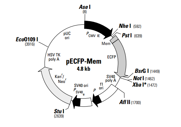
pECFP-Mem encodes a fusion protein consisting of the N-terminal 20 amino acids of neuromodulin, also called GAP-43 (1), and a cyan fluorescent variant of the enhanced green fluorescent protein (EGFP). The neuromodulin fragment contains a signal for posttranslational palmitoylation of cysteines 3 and 4 that targets ECFP to cellular membranes. The ECFP gene contains amino acid substitutions that give ECFP fluorescence excitation (major peak at 433 nm and a minor peak at 453 nm) and emission (major peak at 475 nm and a minor peak at 501 nm) spectra similar to other cyan emission variants (2, 3). These substitutions also enhance the brightness and solubility of the protein (2, 4, 5). In addition to the chromophore mutations, ECFP contains over 190 silent mutations that create an open reading frame comprised almost entirely of preferred human codons (6, 7). Furthermore, upstream sequences flanking the ECFP-Mem fusion protein have been converted to a Kozak consensus translation initiation site (8). These changes increase the translational efficiency of the fusion protein and consequently its expression in mammalian cells.
Expression of ECFP-Mem is driven by the immediate early promoter of CMV (PCMV IE). The vector contains an SV40 origin of replication and a neomycin resistance (Neor) gene for selection in mammalian cells. A bacterial promoter upstream of this cassette (P) expresses kanamycin resistance in E. coli. The vector backbone also provides a pUC19 origin of replication for propagation in E. coli and an f1 origin for single-stranded DNA production.
载体应用
pECFP-Mem can be transfected into mammalian cells using any standard method. If required, stable transformants can be selected using G418 (9). Expression of ECFP-Mem in mammalian cells results in strong labeling of the plasma membrane and allows easy tracking of individual cells in a population. This membrane labeling also permits study of fine cellular processes such as neuronal axons (10), leading edges of migrating cells, filopodia, or microvilli on cell surfaces. pECFP-Mem cannot be used as an exclusive plasma membrane marker because it also partially labels intracellular membranes.

扫一扫 加微信
联系我们
- 客服热线:
0731-84158353 - 订货邮箱:
order@youbio.cn - 技术支持:
tech@youbio.cn  QQ:2605877374